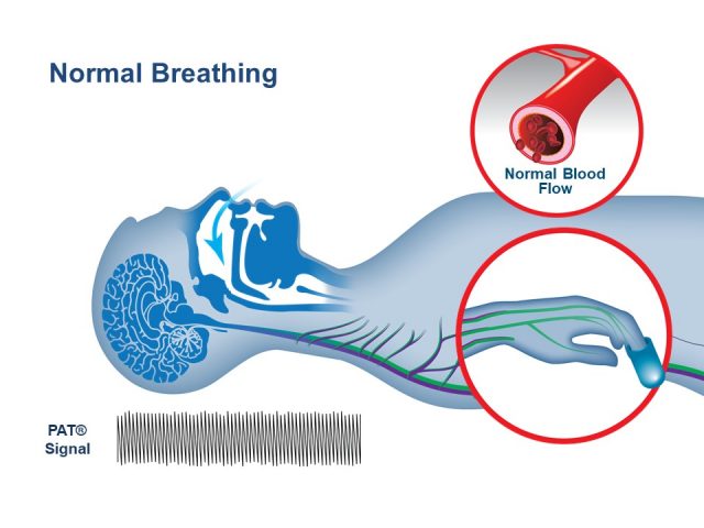
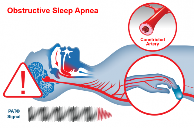
The Peripheral Arterial Tone peripheral arterial tonometry signal is a non-invasive measure of the arterial pulsatile volume changes at the fingertip.
It is a proprietary technology used to identify respiratory events. Using specific signal patterns, the algorithm provides two indices used in determining the degree of sleep apnea, Apnea-Hypopnea Index (AHI) and Respiratory Disturbance Index (RDI). The peripheral arterial tonometry Signal is measured from the fingertip by recording finger arterial pulsatile volume changes since the finger has its unique physiology with;
- High vascular density
- Convenient site for measurement
- Tremendous blood flow variability (1-100ccm/100g/sec)
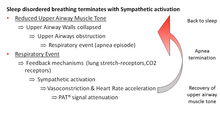
- Reflection of sympathetic nervous system activation, such that terminates any apnea episode
The termination of sleep disordered breathing events is associated with an increase in heart rate, blood pressure, and sympathetic activation. This increase in sympathetic activation results in peripheral vasoconstriction. Peripheral Arterial Tonometry Peripheral Arterial Tonometry measures arterial pulse volume changes in the finger as a result of vasomotion (vasoconstriction and vasodilatation). WatchPAT® home sleep apnea tests use Peripheral Arterial Tonometry signal to detect sleep apnea events. The algorithm analyses the Peripheral Arterial Tonometry signal together with the pulse rate and the oximetry, to accurately diagnose the sleep apnea indices.

Polysomnography (PSG) is considered the “gold standard” for sleep assessment of Obstructive Sleep Apnea (OSA). Numerous validations studies demonstrate a high degree of correlation in RDI, AHI and ODI during simultaneous recording of WatchPAT
and PSG with R= 0.879 (RDI)-0.893 (AHI) and 0.942 (ODI).1


Peripheral Arterial Tonometry Technology FAQ
How does WatchPAT detect Apnea, Hypopnea, and Respiratory Effort Related Arousal (RERA) events?
WatchPATM utilizes Peripheral Arterial Tone Peripheral Arterial Tonometry, a special physiological signal that mirrors changes in the autonomic nervous system (ANS) caused by respiratory disturbances during sleep. The ANS regulates many of our basic functions and it does this without our conscious control. Among its activities are the regulation of blood vessel size and blood pressure, airflow in the lungs, and the heart’s electrical activity and ability to contract. WatchPAT’s automatic algorithm analyzes the Peripheral Arterial Tonometry signal amplitude along with the heart rate and oxygen saturation to identify and classify breathing problems while you sleep. Using specific signal patterns, the algorithm provides two indices that allow a diagnosis of sleep apnea: • AHI (Apnea/Hypopnea Index), which is an index used to calculate sleep apnea severity based on the total number of complete cessations (apneas) and partial obstructions (hypopneas) of breathing per hour of sleep. • RDI (Respiratory Disturbance Index) is used to assess the severity of sleep apnea by measuring respiratory efforts, or RERAs (Respiratory Effort Related Arousals). A RERA is an arousal from sleep that follows 10 seconds or more of increased respiratory effort but does not meet the criteria for apnea or hypopnea. The snoring sensor enables the clinician to determine if the respiratory events are obstructive and the body position sensor enables the clinician to determine if there is a positional component to the sleep apnea
How does WatchPAT detect rapid eye movement (REM) from the finger?
Rapid eye movement (REM) sleep is associated with considerable attenuation of the Peripheral Arterial Tonometry signal and physiology coupled with specific variations in the Peripheral Arterial Tonometry amplitude and rate. Based on this specific variability in the Peripheral Arterial Tonometry and pulse rate signals, REM sleep stage is differentiated from non-REM sleep. In addition, it is differentiated from the wake state by WatchPAT’s advanced actigraphy algorithms. This is clinically important to prevent under-treatment of apnea if predominantly REM-related.
How does the WatchPAT home sleep apnea test detect sleep architecture?
WatchPAT’s sleep/wake detection is based on data recorded by the built-in actigraph. The propriety software’s automatic actigraph algorithm discriminates between sleep and wake states in normal subjects and those with Obstructive Sleep Apnea (OSA). This algorithm makes WatchPAT’s home sleep apnea test (HSAT) superior to all other actigraph devices—most of them fail when used with OSA subjects. WatchPAT’s sleep/wake algorithm has been validated and published in peer-reviewed journals. The results show very good agreement between actigraphy and PSG in determining sleep efficiency, total sleep time, and sleep latency (agreement 86% in normal subjects, 86%-mild OSA, 84%-moderate OSA, 80% severe OSA).
How does WatchPAT differentiate between respiratory arousals and other events like Periodic Limb Movement (PLM) related arousals and spontaneous arousals?
The algorithms differentiate between respiratory related arousals and Periodic Limb Movement (PLM) related or spontaneous arousals via detection of specific patterns in the Peripheral Arterial Tonometry signal coupled with the presence or absence of unique dynamics of the pulse oximetry signal.
